Contents

|
|
Amount |
Cat. No. |
Introduction |
Storage |
|
iMSC-luc-GFP(P2) |
1×10⁶ |
RC02011 |
Insertion of Luc and GFP gene fragments at ROSA26 locus, expressing Luciferase and green fluorescence protein (nuclear) |
Liquid nitrogen |
|
iMSC-Anterase2 (P2) |
1×10⁶ |
RC02012 |
Insertion of Anterase2 gene fragments at ROSA26 locus, expressing Anterase2 |
|
|
ucMSC-luc-GFP(P3) |
1×10⁶ |
RC02013 |
ucMSC,insertion of Luc and GFP
gene fragments at random,
expressing Luciferase and green fluorescence protein (nuclear) |
|
|
ucMSC-Anterase2(P3) |
1×10⁶ |
RC02014 |
ucMSC,insertion of Anterase2 gene fragments, expressing Anterase2 |
- Overview
- Citation
-
Fluorescent protein labeled MSC is generated by insertion of reporter into the target sites using gene editing technology. These constructed MSC can efficiently express Luciferase and GFP, proliferate stably, with normal karyotype and specific cell surface factors (CD73+ / CD90+ / CD105+, CD14- / CD34- / CD45- / CD79α- / HLA-DR-) retained. These cells can realize in vivo imaging of animals after cell transplantation. iMSC-Luc-GFP can be applied in various in vitro experiments, drug screening and safety evaluation, as well as cell transplantation in disease animal model.
Anhui Shownin Biotechnologies Co., Ltd. has a variety of different MSC cell lines, please call 400-888-3920 for detailed consultation.
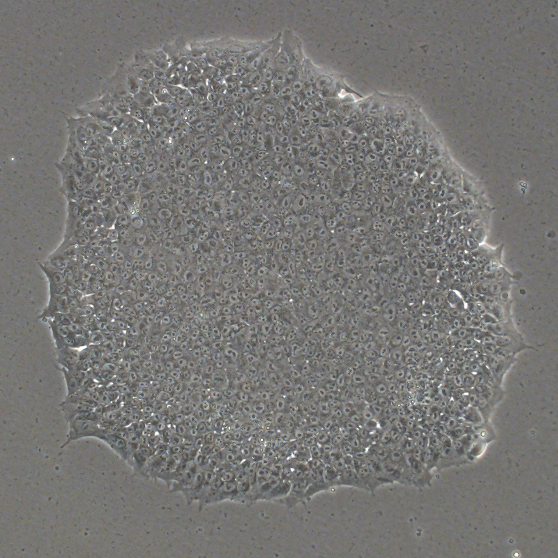
Cell morphology

The morphorlogy of iMSC-Luc-GFP cultured at day 3 after passaging (P4) in ncMission hMSC Medium of 8,000 cells/cm2 at seeding.
A :iMSC B : iMSC-luc-GFP C : Fluorescence image of iMSC-luc-GFP cultured at day 3. Scale bar, 200 μm.
Small animal live imaging display

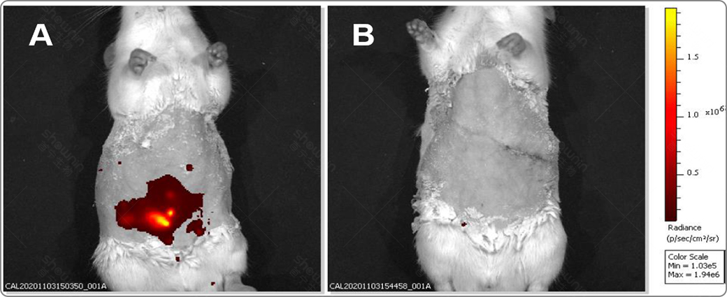
Imaging of rats with ucMSC-Anterase2 cell line.
Figure A is the experimental group and Figure B is the control group. ucMSC-Anterase2 signal is highly expressed, and in vivo imaging can be efficiently achieved in rats with thick cell walls.
Related products


